(Source: https://unsplash.com/photos/f2aDTqfnqfE)
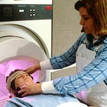
Medical imaging creates images of internal parts of the human body for diagnosis and clinical investigation purposes. Various imaging techniques and equipment are used for this purpose. Some of the widely used imaging techniques include X-ray radiography, magnetic resonance imaging, endoscopy, tactile imaging, medical photography, functional imaging techniques such as Positron Emission Tomography (PET) and Single-Photon Emission Computed Tomography (SPECT). Physicians have started adopting these different techniques to diagnose diseases and plan their treatment accurately.
Further, these imaging techniques are generating high-resolution images. Analysis of which can be automated through computational techniques. The goal of automated image processing is twofold. The first goal is to measure the features on medical images, e.g., helping radiologist obtain measurements from medical images (e.g., area or volume). The second goal is to make a diagnosis from these measures and provided features. Such automated techniques help radiologists with their diagnosis procedure for accuracy and efficiency.
The field of image processing and analysis heavily depends on human expertise and so prone to error. Many developing countries face a shortage of human knowledge. Hence the timely and accurate diagnosis of medical images become the main concern for health care delivery even when the healthcare system is strengthened with proper medical equipment.
Hence one way to address this challenge is to adopt the advances in digital image processing to analyse medical images, interpret the image content and recommend a treatment schedule. The majority of the imaging equipment listed earlier in this section produce images in digital format. Hence they can be processed by applying image analytical functions developed in digital image processing. These analytical functions are:
Filtering: To reduce noise and blur.
Segmentation Identify structures within the image.
Quantification: Extract useful information from the image.
Enhancement or Reconstruction Prepare the image for visualization.
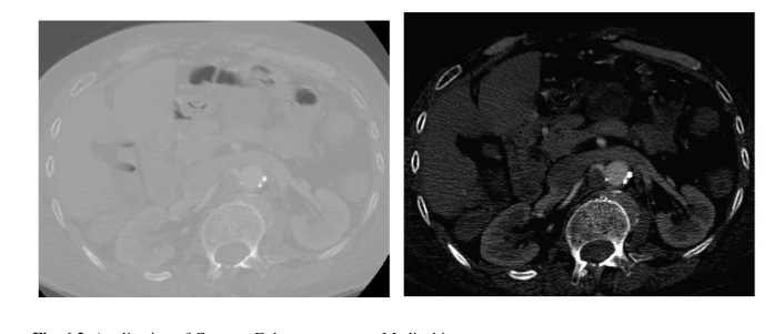
The figure shows the applications of contrast enhancement function on a medical image. These primitive functions are used to realize high-level tasks of object detection and classification. For example, outgrown tissue or abnormal growth of a tissue called a tumour can be detected. Further, the detected tumour can be classified as a malignant or benign tumour.
Machine learning and deep learning techniques are used to realize these high-level tasks of object detection and classification. Machine learning techniques such as decision trees, Naive Bayes classifiers and Support Vector Machines are preferred when pre-defined features can either detect an object from a given image or classify the image. At the same time, techniques of deep learning such as Convolutional Neural Network, Recurrent Neural Network are preferred when images are high-dimensional, and features used for classifying images are not well defined.
So far the machine learning and deep learning-based techniques have been used for the diagnosis of diseases like Diabetic Retinopathy, Histological and Microscopical Elements Detection, Gastrointestinal (GI) Diseases Detection, Cardiac Imaging, Tumor Detection, Alzheimer’s and Parkinson’s Diseases Detection.
Development and deployment of solutions based on medical image processing for healthcare delivery face many issues and challenges. The first challenge is the availability of disease-specific validated open dataset to develop machine learning-based classification and object detection techniques. The second most important challenge is following data privacy standards in the health care industry which vary from region to region.


